Cerebellum and Cisterna Magna
Reason for evaluation: exclude two main entities:
- Dandy Walker Malformation
- Chiari Malformation.
When evaluating the cisterna magna, you basically need to know if it’s either too big or too small. You can usually identify this with a simple “eye test.” In fact, I do not routinely measure the cisterna magna.
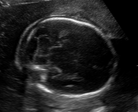
Figure 4.1. Normal cisterna magna and cerebellum. Burn this picture into your mind as what normal looks like. The cisterna magna measures < 10 mm, and the cerebellum maintains a nice normal shape and the central portion of the cerebellum (the vermis) is intact.
If the cisterna magna is too large (>10 mm), you need to be concerned about a Dandy Walker malformation which is due to agenesis of the cerebellar vermis (the central portion of the cerebellum).
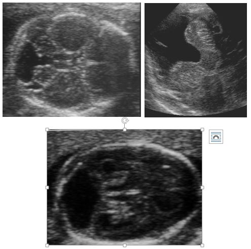
Figure 4.2. All three images show varying degrees of vermian agenesis. In every case, the cisterna magna is clearly large and > 10 mm. The cisterna magna also communicates with the 4th ventricle.
You will notice that in the three cases in figure 4.2, the cisterna magna is visually abnormal and all measure larger than 10 mm and the cerebellar vermis is absent.
If the cisterna magna is large, and the vermis is present, then you are most likely either dealing with a mega cisterna magna or an arachnoid cyst. These two entities tend to be of lesser clinical significance. In fact, they are often incidentally found in adults on routine brain imaging for another reason.
An important observation when faced with this situation is to identify the 4th ventricle as a fluid-filled triangle. In the above three pictures, the 4th ventricle is wide open to the cisterna magna. In the below picture (Figure 4.3) of an arachnoid cyst, the 4th ventricle can still be identified.
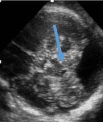
Figure 4.3. There cisterna magna is clearly large. Our next step is to attempt to identify the 4th ventricle (blue arrow). If the 4th ventricle does not communicate with the cisterna magna, then you are dealing with a mega cisterna magna or arachnoid cyst.
If you can’t find a cisterna magna to even measure (i.e. it is completely effaced) then you are most likely dealing with a Chiari malformation. This shape of the cerebellum is referred to as the banana sign. You can see that in the picture below (Figure 4.4), the cerebellum looks is banana shaped and there is no discernible cisterna magna. Unfortunately, it is not always this pretty, so if you are having trouble even seeing the cisterna magna, you should be suspicious. Of course, Chiari malformations are also associated with ventriculomegaly and a distal spinal dysraphism.
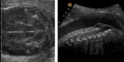
Figure 4.4. There is not visible cisterna magna. The cerebellum has a “banana” configuration, consistent with a Chiari malformation and should prompt a detailed evaluation of the distal spine for a myelomeningocele.
Figure 4.5. The still image in Figure 4.4 shows a near perfect “banana sign” but often times, when there is a Chiari malformation, due to the small cisterna magna, crowding of the cerebellum and shadowing from the bones, it is difficult to see well. Thus, you must maintain a high index of suspicion if you are not able to get a good view of the cisterna magna.